Cells
S1 lessons on living things, microscopes, plant and animal cells, specialised cells, microbes and body defences. Choose a lesson to open it.
- Lesson 1Living or Non-Living?
- Lesson 2Using a Microscope
- Lesson 3Cell Structures
- Lesson 4Consolidation
- Lesson 5Onion Cell Slide
- Lesson 6Cheek Cell Slide
- Lesson 7Specialised Cells
- Lesson 8Microbes
- Lesson 9Researching Microbes
- Lesson 10Microbial Growth and Graph Skills
- Lesson 11Body Defence
- Lesson 12Vaccines (coming soon)
Lesson 1 - Living or Non Living?
Learning Intentions
- Know how to recognise living things.
- What all living things are made up of.
- Name the parts of a microscope.
Living or Non Living?
Task 1
Sort the cards into two groups: living and non-living.
Task 2
Answer the following questions:
- How did you decide which pile to put the cards into?
- What do the cards in the living pile have in common?
- What makes them different from the non-living pile?
- What can living things do that non living things cannot do?
Task 3
Watch the video below.

Notes - The seven processes of life
- M - movement
- R - respiration
- S - sensitivity
- G - growth
- R - reproduction
- E - excretion
- N - nutrition
Every living thing is made up of cells.
Most cells are too small to see without a microscope. A microscope is used to magnify objects (make them look bigger).
Cells
Do you know what types of cells are in the following images?
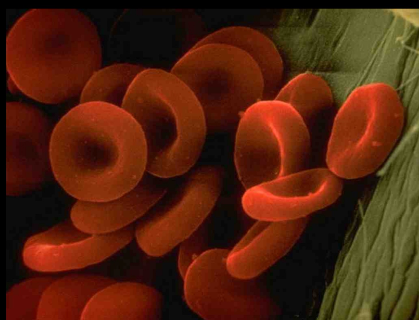
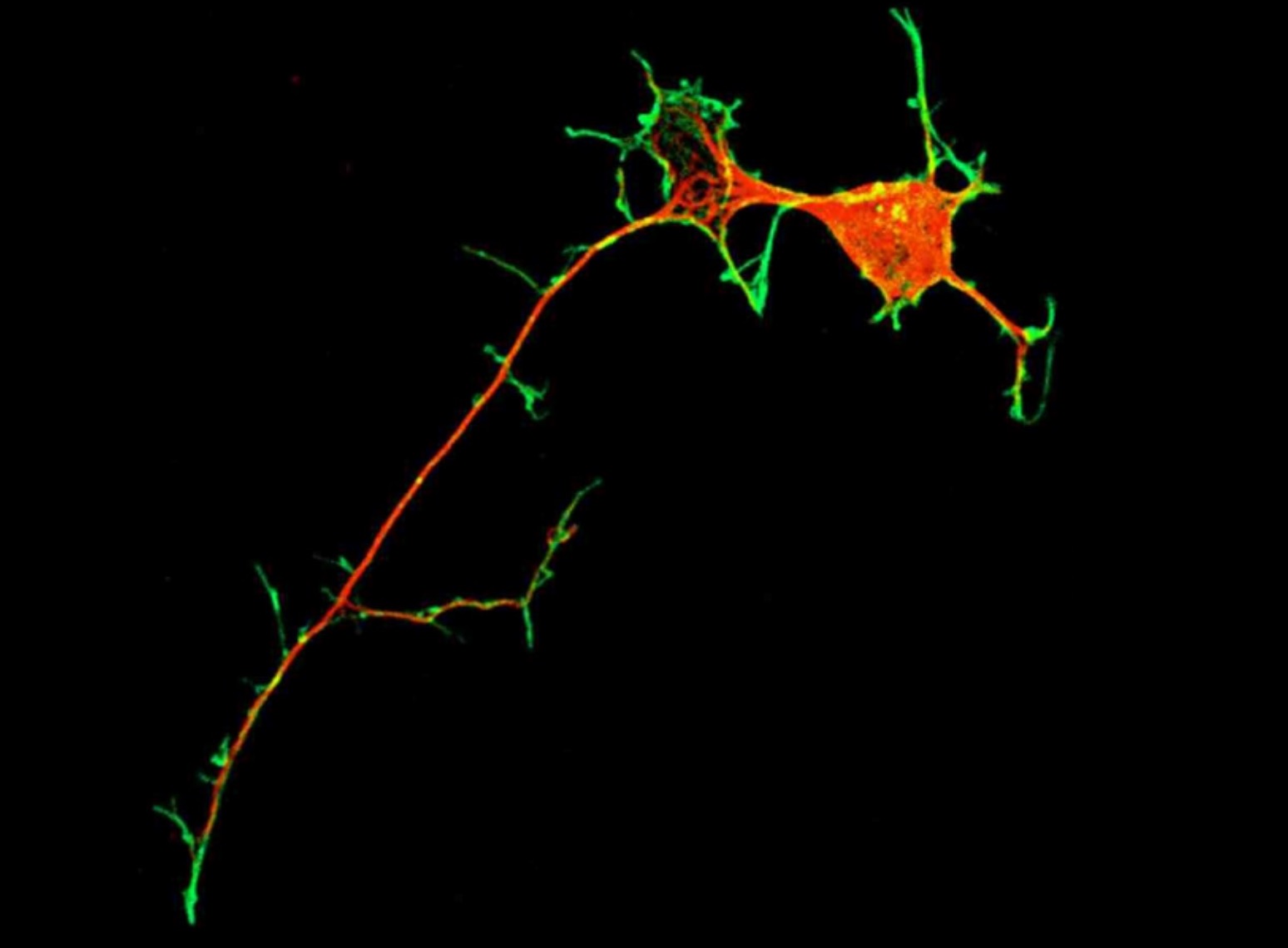
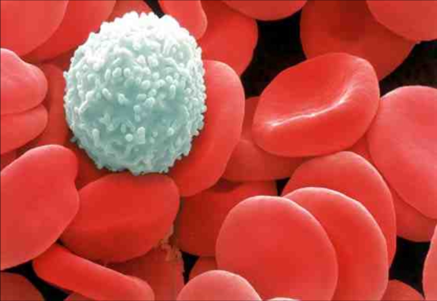


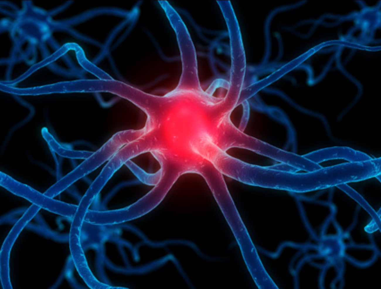
The Microscope
Collect a microscope.
Carry the microscope by the handle, with one hand underneath the base.
When carrying the microscope it should be the only thing you have in your hands.
Working in pairs, use bluetack to add labels to each part of the microscope.
OR
Drag the labels to the correct parts of the diagram below.
Have your work checked before filling in the labels on your microscope diagram.
Stage
Fine Focusing Dial
Rough Focusing Dial
Mirror
Eyepiece

Lesson 2 - Using a Microscope
Learning Intentions
- By the end of this lesson, I will be able to use a microscope safely.
Using a Microscope
- Watch your teacher demonstrate the correct use of a microscope.
- Work in pairs to set up a microscope.
- View a slide with a sample cut from newspaper on it.
Note - Looking through a Microscope
When an object (thing you are looking at) is placed under the microscope its image (what we see) is u_____ d___ and b___ t_ f____.
Microscope Magnification
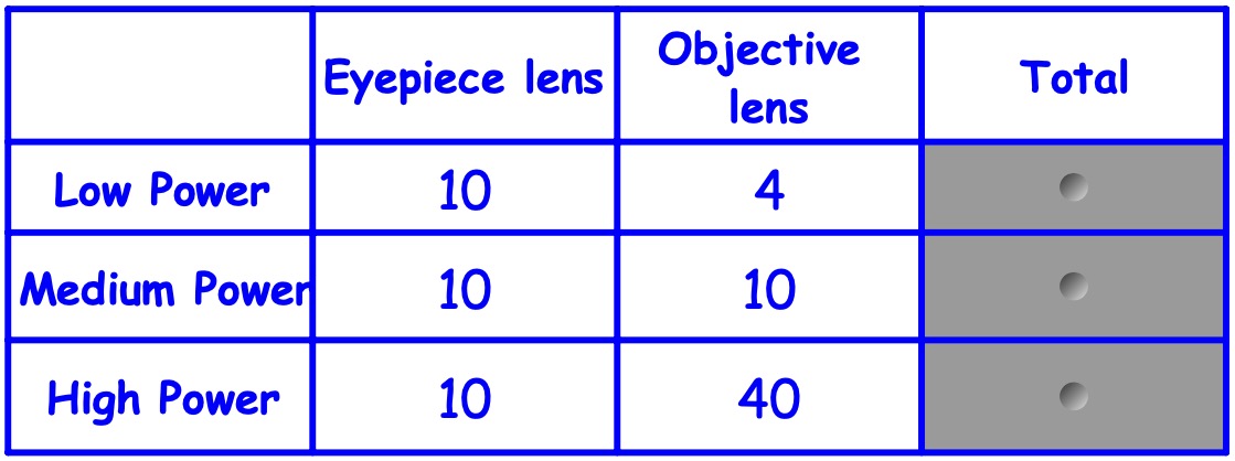
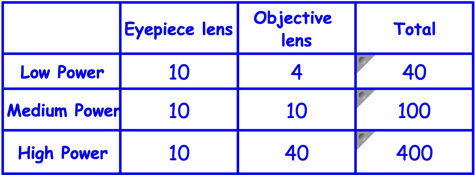
The number of times an object is made bigger is called the magnification.
This is worked out by:
Eyepiece Lens 'strength' x Objective Lens 'strength'
Look at the number written on the eyepiece lens and on the objective lenses. Then copy and complete the table:
Using a Microscope
- View a variety of different slides under the microscope.
- Draw each thing you look at (approx. 1/3rd of a page in size).
- Make sure that each one of your drawings has a title, border and the magnification is written next to it.
- Try looking at the screen of your phone.
- Be careful not to crack your screen!
Lesson 3 - Cell Structures
Learning Intentions
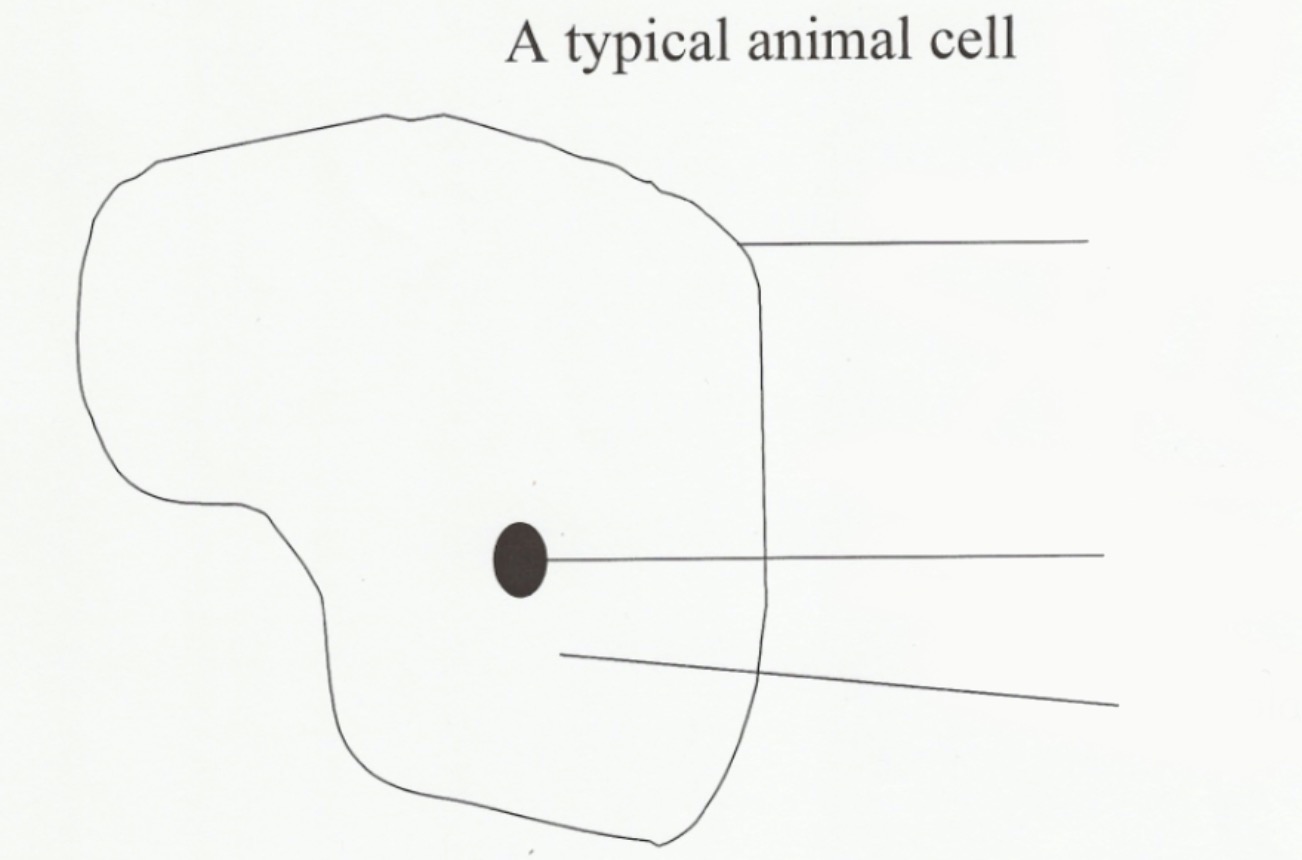
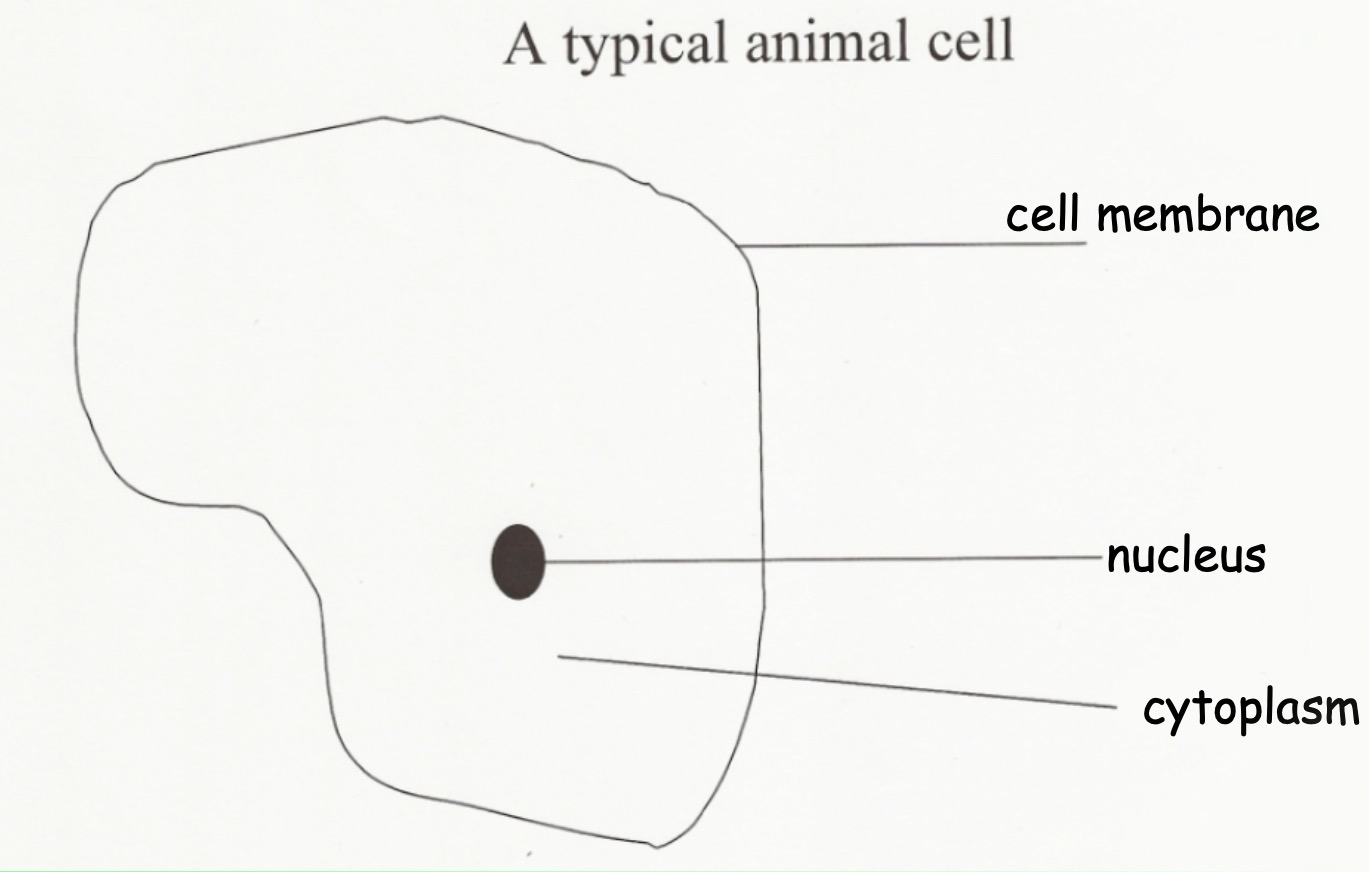
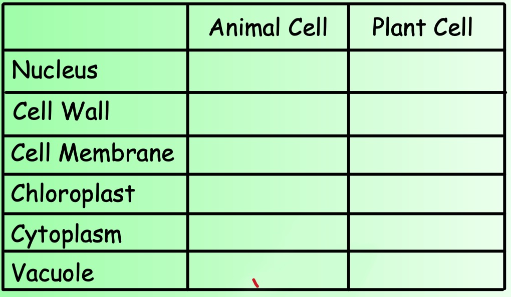
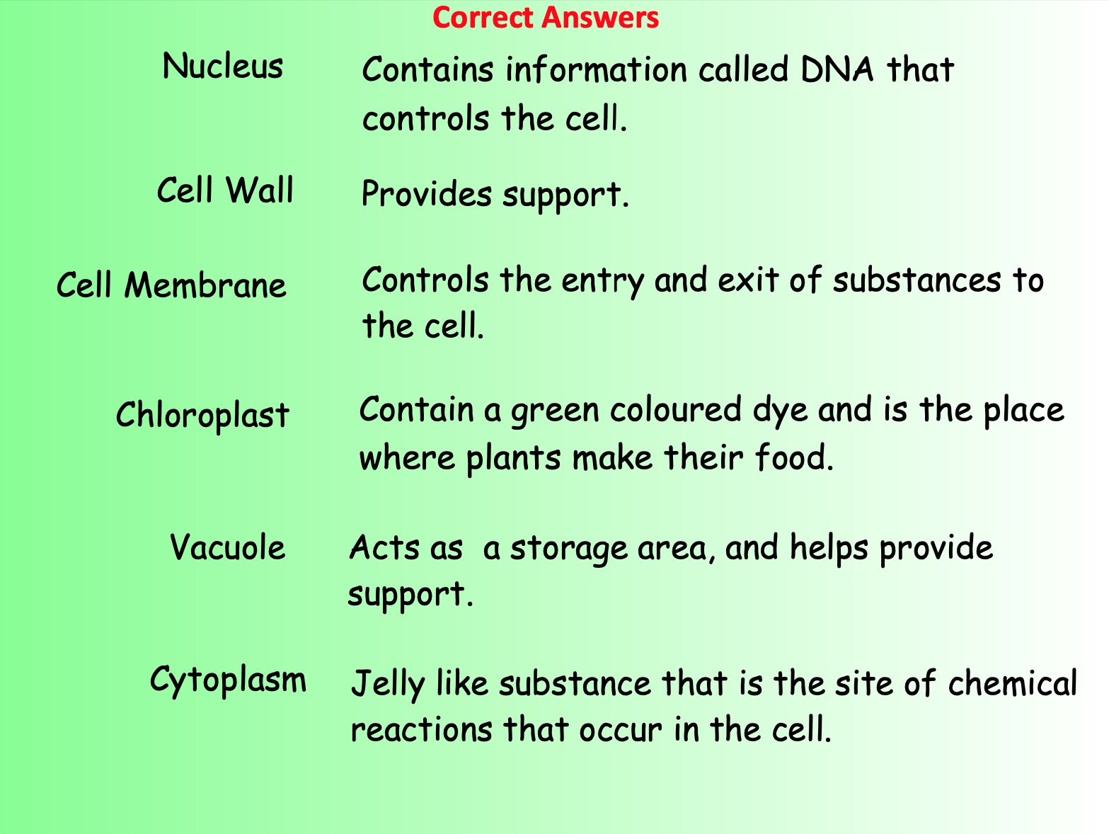
- Nucleus, Cytoplasm and Cell Membrane in an animal cell
- Nucleus, Cell Wall, Chloroplast, Cytoplasm, Vacuole in a plant cell.
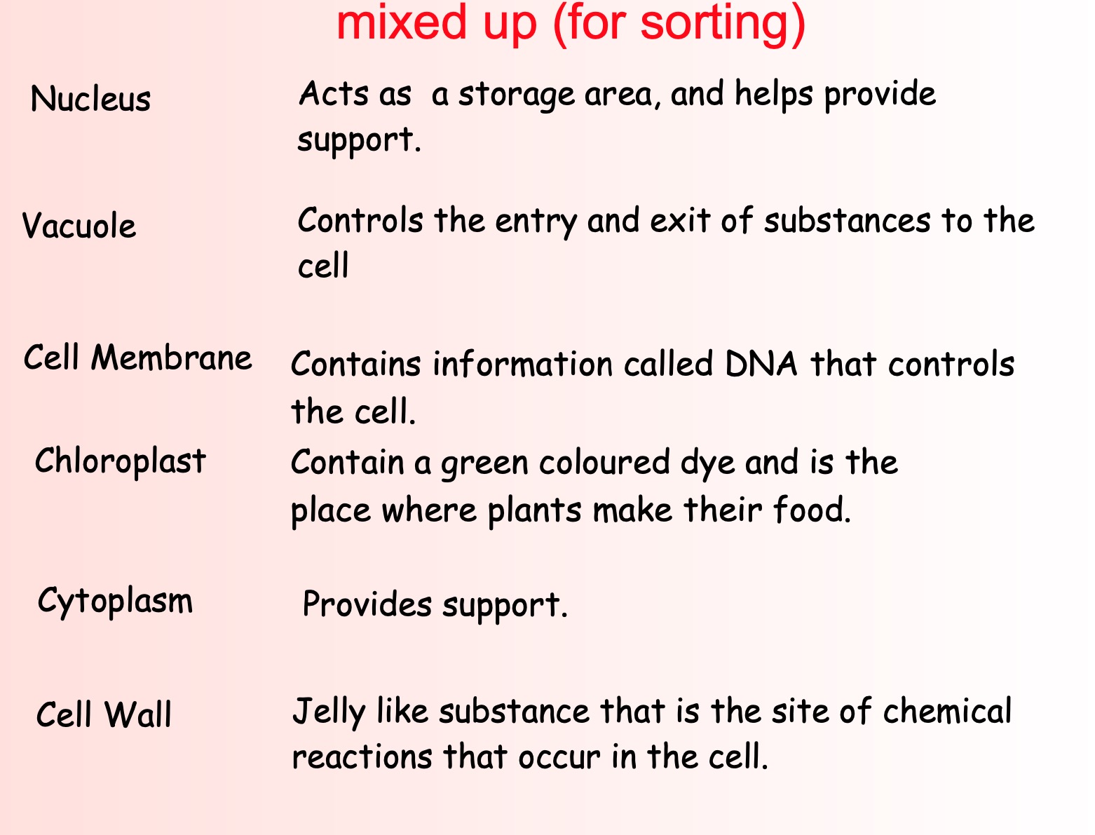
- And describe the function of each part.
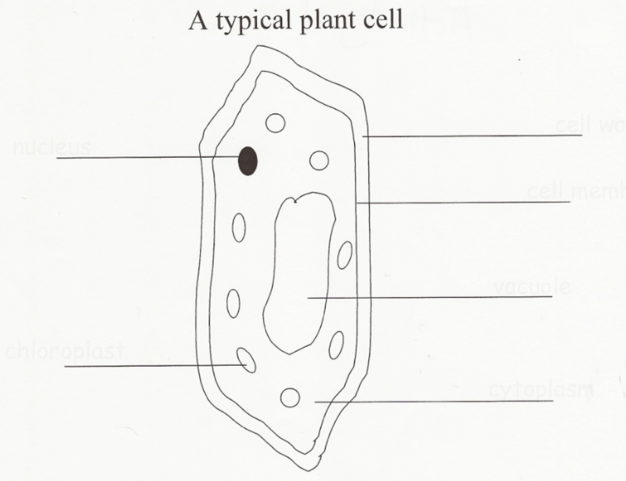
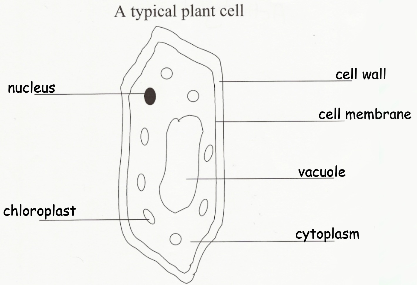
Animal vs Plant
Cell part functions
Lesson 4 - Consolidation
Learning Intentions
- Consolidate learning on cells
Cells Consolidation
Watch the videos below on what we have found out so far about cells.
Cells - Graphic Organiser
Collect and complete the cells graphic organiser worksheet.
Lesson 5 - Making an Onion Cell Slide
Learning Intentions
- I will be able to use a microscope, and make a slide.
Activity - Making an Onion Cell Slide
Collect and glue in the "Making a Slide" handout and read it carefully.
Watch your teacher demonstrate how to make an onion cell slide.
Make your own onion cell slide. Use the different lenses on your microscope to look at your slide.
What you should have seen

What was the purpose of adding iodine solution to your slide?
Note - Staining
A stain is used to make the cell structures stand out. The iodine solution will stain the nucleus of the onion skin cells.
Lesson 6 - Making a Cheek Cell Slide
Learning Intention
- I will be able to use a microscope, and make a slide.
Making a cheek cell slide
Look back over your hand out from last lesson on "Making a Slide".
Watch your teacher demonstrate how to make a cheek cell slide.
Make your own cheek cell slide. Use the different lenses on your microscope to look at your slide.
What you should have seen
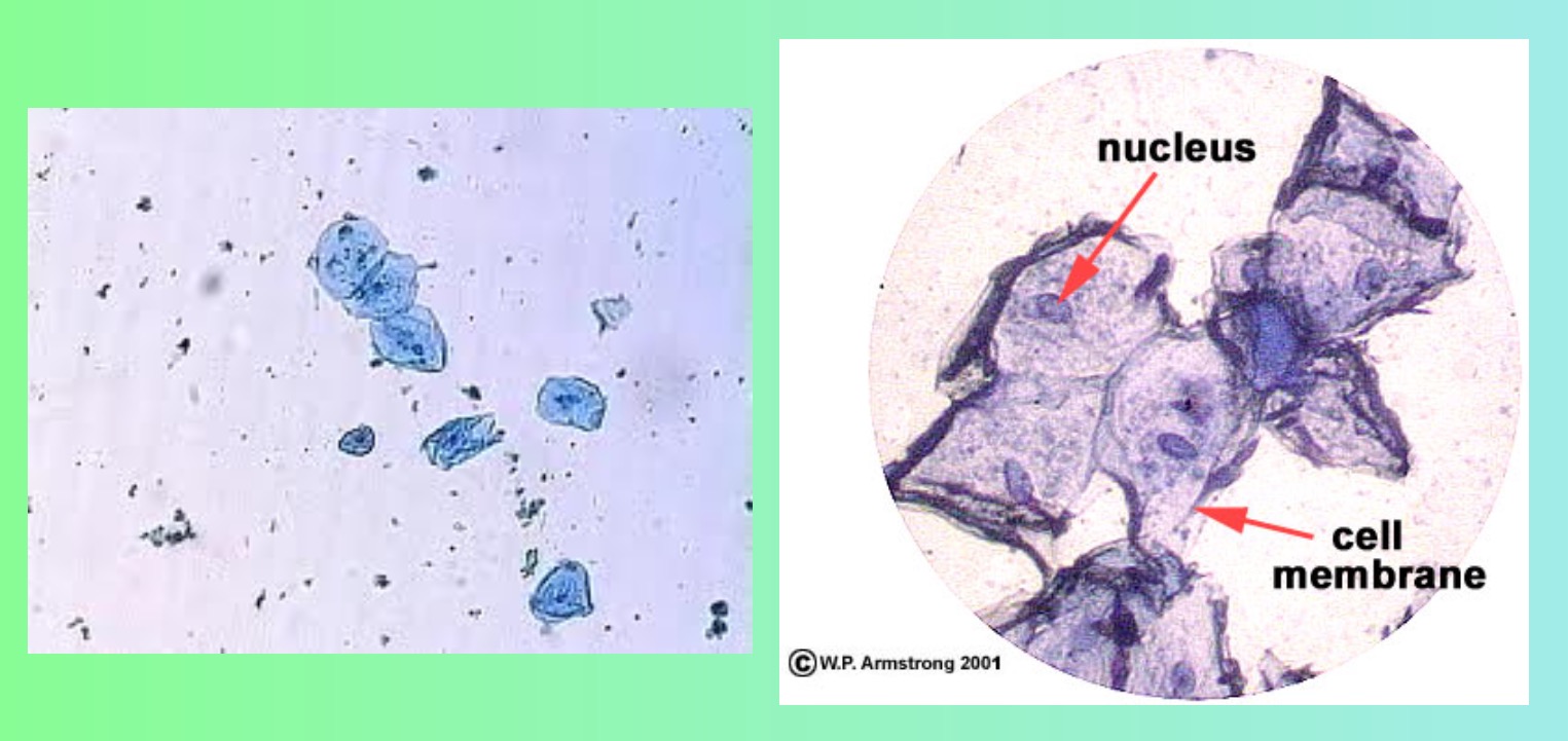
Draw your cheek cells, including:
- a label for the nucleus
- the magnification
Notes - Cheek Cell Slides
Cheek cells are flat in order to provide a lining that will protect our bodies from things like bacteria.
Cheek cells are said to be specialised because their structure allows them to do a special job.
Lesson 7 - Specialised Cells
Learning Intentions
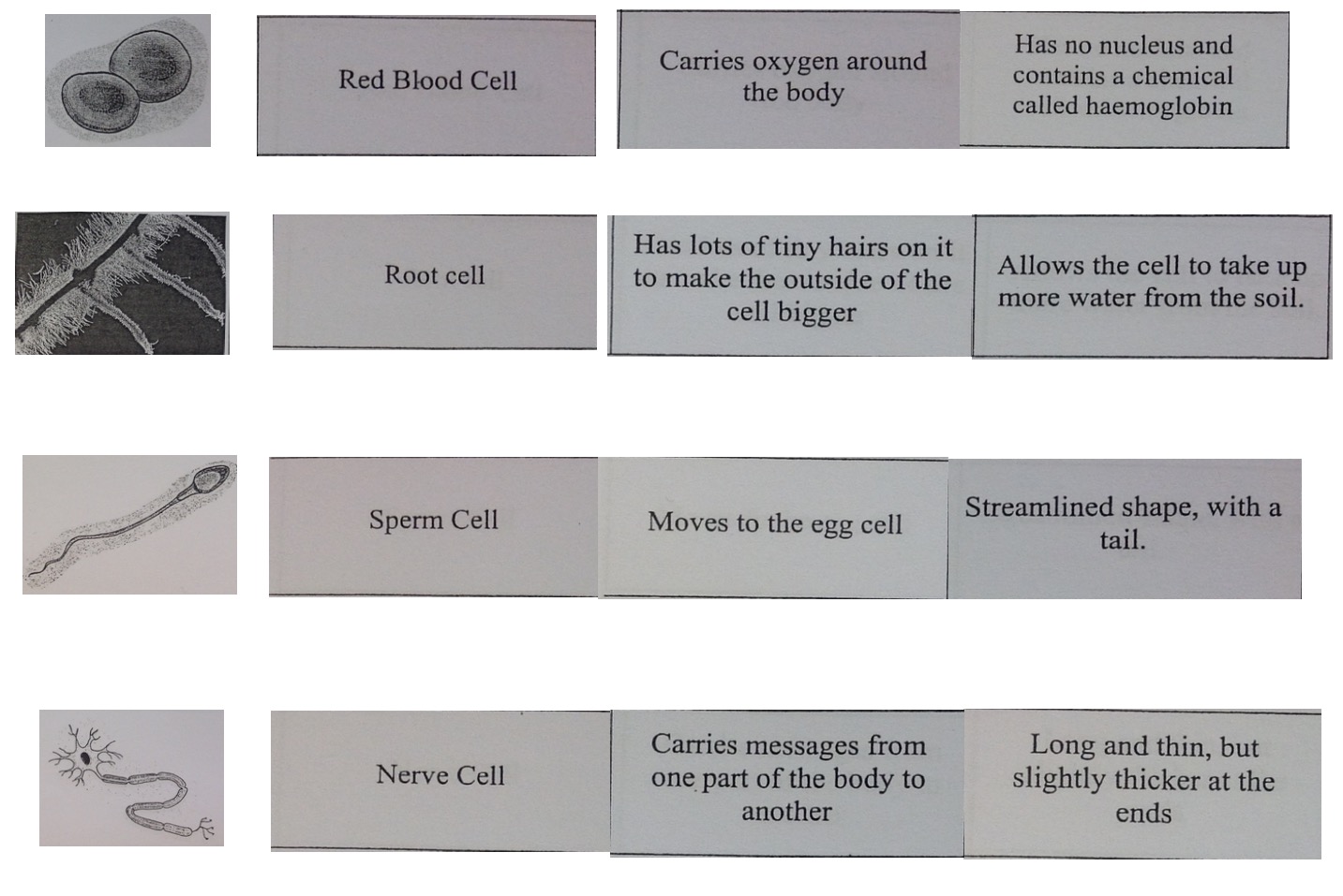
- I should be able to match pictures of specialised cells with information on their function.
Note - Specialised Cells
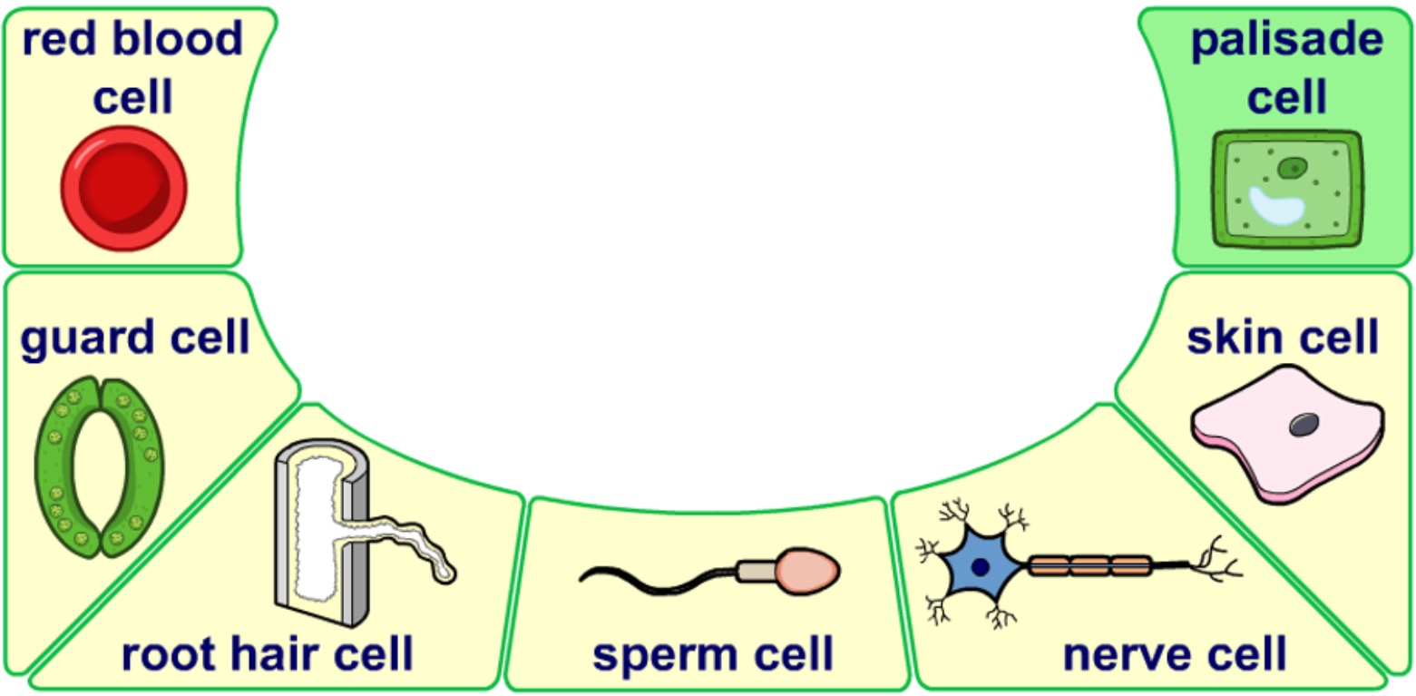
Plant and animal cells are often different shapes and may not have all the usual cell structures. This allows them to carry out different jobs.
For example:
- Palisade cells contain many chloroplasts to trap light.
- Xylem cells hollow themselves out then die - to form tubes.
- Cheek cells are flat to form a protective lining.
Try to match up the pictures of the cells with their name. Think about their structure. Does this help them to carry out their function?

Activity - Specialised Cells
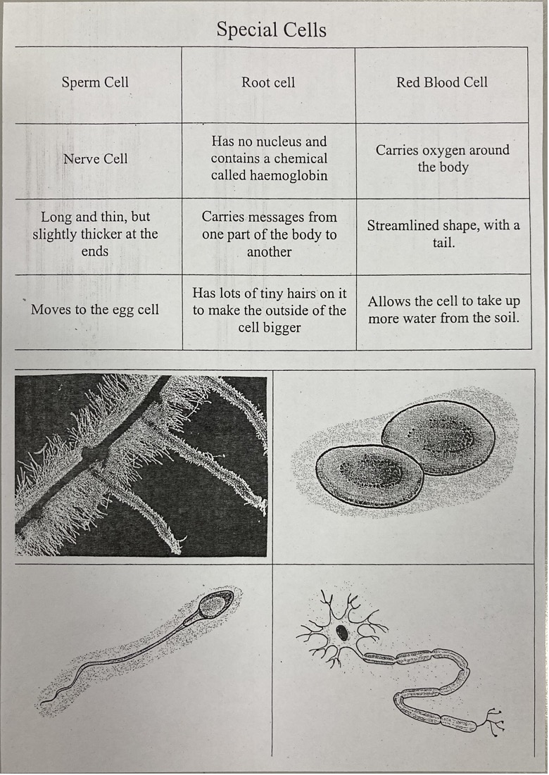
Collect the sheet Special Cells.
- Cut out the cell diagrams and cell names.
- Match them with the statements
- Stick into your jotter in correct order.
Lesson 8 - Microbes
Learning Intentions
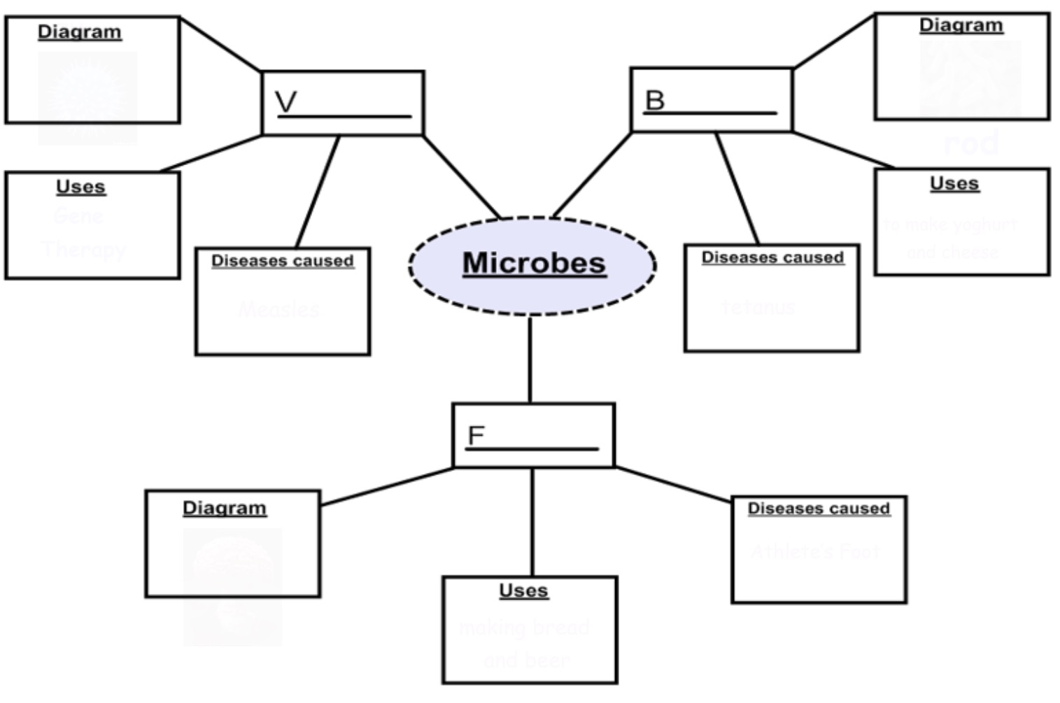
- State that bacteria, fungi and viruses are microbes.
- Describe some of the uses of microbes.
Variety of Microbes
Make sure that you have a copy of the Microbes spider diagram below. Fill it in as you read the following information on microbes.
Fungi
Diagram

Uses
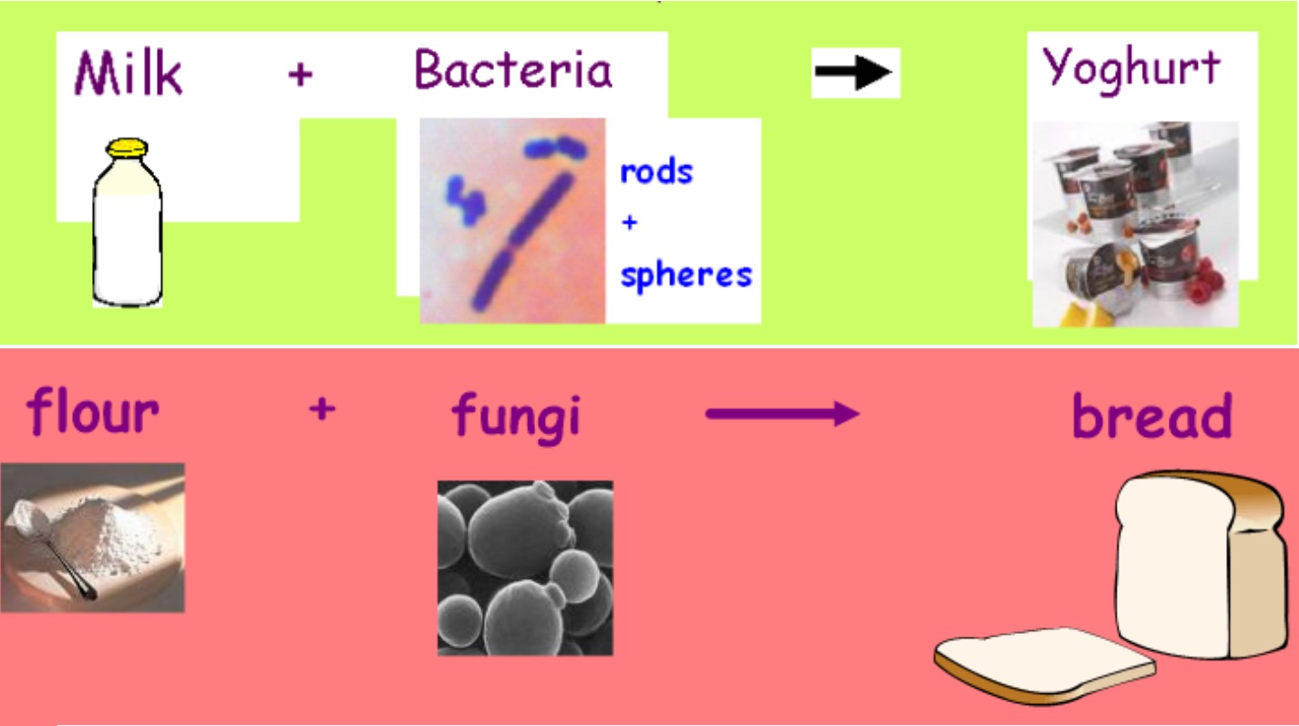
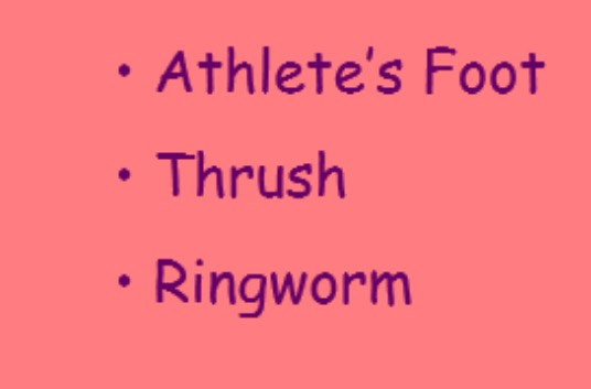
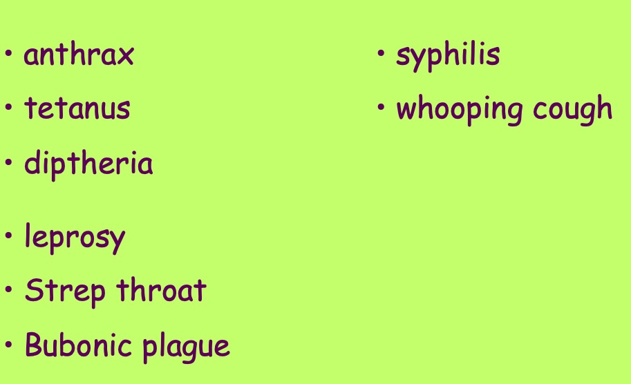
Diseases
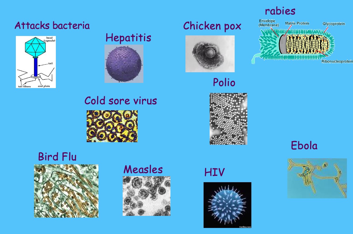
Viruses
Uses
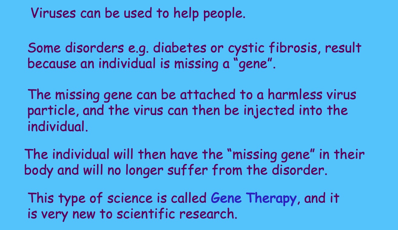
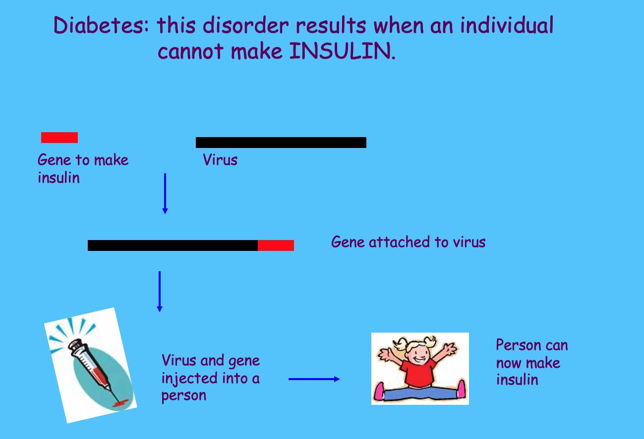
Diagram

Diseases
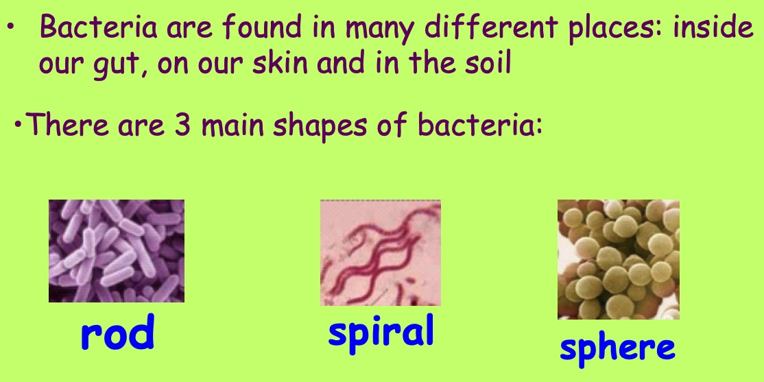
Bacteria
Uses

Diagram
Diseases
Lesson 9 - Researching Microbes
Learning Intentions
- Diagrams of the microbes
- What they're used for
- Any diseases they cause
- Other interesting facts
Activity - Researching Microbes
In this task you will be choosing a particular microbe (virus, bacteria, protozoa or fungi) to research. Watch the videos below and follow the links below to help you decide which microbe you would like to research.
Find out some information about your chosen microbe. Use the 'Microbe Investigation' worksheet below to help you.
Once you have found out some information about your chosen microbe you will present it to the rest of the class.
Lesson 10 - Microbial Growth and Graph Skills
Learning Intentions
- I will be able to explain how the growth of microbes can be controlled.
Starter
Thinking about what you have learned about microbes from previous lessons, brainstorm with your partner what you think might be the best conditions for microbes to grow.
Make a list of three things that would let microbes grow well.
Notes - Microbial Growth
Microbes are often grown in laboratories in fermenters, flasks and on agar plates.
To grow well, microbes need a supply of nutrients (e.g. sugar) and a suitable (e.g. 20-40oC) temperature. Scientists can control the growth of microbes by altering the nutrient supply or the temperature.
Microbes can be killed by :
- radiation e.g. UV and gamma rays
- chemicals such as alcohol or disinfectant
- very high temperatures e.g. 120oC.
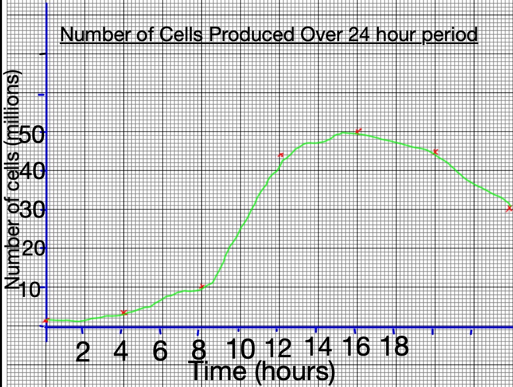
Graph Skills
Collect the sheet Yeast Industries: Yeast and Bread Making and a blank sheet of graph paper.
Use this step by step guide to help you to draw a line graph
Step 1
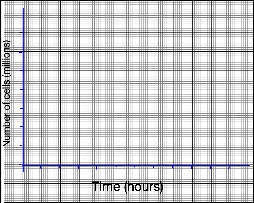
Draw the x and y axes on your graph.

Step 2
Label the axes.
Step 3
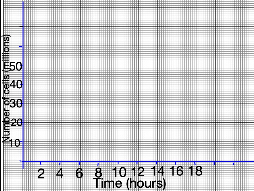
Decide on a scale for both axes. The scale should increase in regular intervals.
Step 4
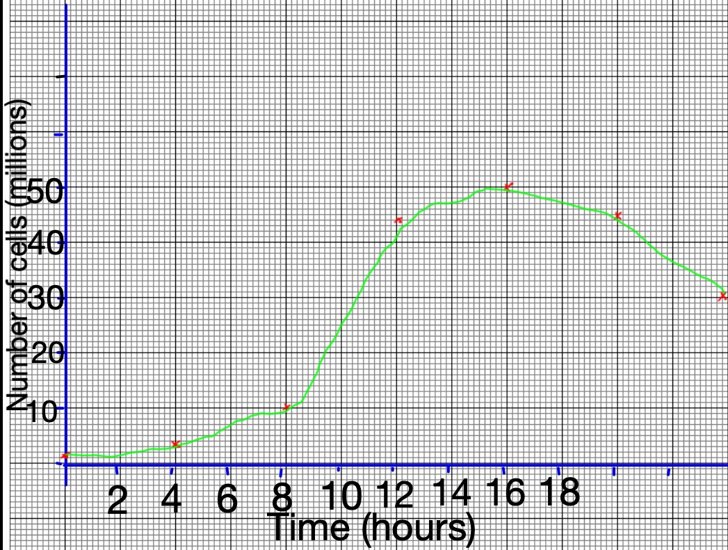
Use the data from the table on the worksheet to plot the points. Once you have done this, draw a line of best fit.
Step 5
Give your graph a meaningful title
Lesson 11 - Body Defence
Learning Intentions
- I will know about the three ways the body can be defended against microbes: external, internal and artificial defences.
Notes - Body Defences
As a minimum, note the words in blue.
(1) External Defences
- Sticky mucus in the windpipe stops microbes getting into your lungs.
- Your skin is a barrier, it stops microbes getting into your body.
- Acid in your stomach kills microbes that you swallow.
(2) Internal Defences
- White blood cells engulf (surround) and destroy microbes in the blood.
- Antibodies also destroy microbes in the blood.
This is your IMMUNE SYSTEM.
(3) Artificial Defence
- Vaccinations e.g. HPV, MMR